A Personal Journey: The Urgent Need for Better Breast Cancer Screening
By Sheila Mikhail
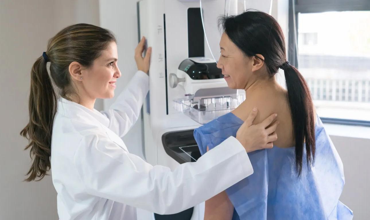
Breast cancer was not the first thing on my mind last November when I noticed a small dimple on my left breast. Earlier that year, I received a "clean" mammogram report from Duke, utilizing 3D tomosynthesis (DBT), and I have no family history of breast cancer. Having just celebrated my 56th birthday and gained some weight during my transition into deeper menopause, I initially dismissed the dimple as cellulite. However, after urging from my family, I decided to request a diagnostic mammogram.

During the workup, a diagnostic mammogram was performed on both breasts. The results revealed a sizable tumor on the left side. Ten days later, I received the confirmation: I had breast cancer. During my visit with the oncologist, I inquired about the possibility of having breast cancer on the right side and requested additional imaging. The oncologist pushed back, stating that it was "not the standard of care" and that “insurance would not pay.” Frustrated by my insistence, she remarked that I was making a “ruckus.” After I reiterated my willingness to self-pay, she finally ordered CT and breast MRI tests.
A week later, the breast MRI confirmed the left-side tumor, which measured 2.8 cm upon removal, and identified a second sizable tumor on the right side, extending nearly 6 cm. This newly detected tumor had gone unnoticed by both the DBT and the diagnostic mammogram. Oncologists believe these tumors were slow-growing and had likely been developing for up to a decade before their discovery.
I’ve had mammograms for many years without issues—except for one instance. My records show I was called back for additional testing in 2013 when something suspicious was found in both breasts. Although I underwent a diagnostic mammogram, no biopsy was performed, and the findings were dismissed as benign. Given that I had been informed I had heterogeneously dense breasts, I consulted my Duke primary care physician (PCP) and my UNC gynecologist (GYN) about whether I needed further testing. They assured me that, due to my lack of family history, 3D tomosynthesis would suffice. I also attended annual physicals with my PCP and GYN, during which my breasts were examined. I conducted monthly self-examinations but felt they were not helpful due to the fibrotic nature of my breasts.
Almost two months after the identification of my first tumor, I underwent surgery to remove both tumors, followed by 66 radiation treatments. I did not receive chemotherapy, partly because my type of breast cancer— invasive lobular carcinoma—does not respond well to it. Interestingly, this form of cancer, which represents 15% of breast cancers, often lacks a palpable lump. UNC provided exceptional cancer care, and I was informed that my prognosis was good; I had caught it just in time to prevent metastasis.
According to the National Cancer Institute (NCI), the mortality reduction through screening is significantly lower in women with dense breasts, as tumors are often not readily detectable by mammography. By making more sensitive imaging modalities accessible to women with dense breasts, we can potentially catch more cases of breast cancer early and save lives.
Breast cancer affects one in every eight women, and its prevalence is on the rise. Despite advancements in treatment, the American Cancer Society (ACS) predicts that 43,170 women in the U.S. will die of breast cancer in 2023—an avoidable statistic. If caught early, breast cancer is almost 100% survivable; however, if detected late, the ACS estimates only a 30% chance of surviving for five years. Based on NCI data, while mammography is effective for 80% of the population, it fails to adequately serve the remaining 20%, which equates to approximately 9,300 women in North Carolina each year.
Mammography is less effective at detecting tumors in women with dense breasts, missing about 50% of tumors, according to a 2017 study published in JAMA Oncology. This is due to dense breast tissue appearing white on mammograms, just like cancer. Essentially, it’s akin to searching for a snowball in a snowstorm. This lack of visibility is concerning, especially since women with dense breasts are at a higher risk of developing breast cancer. A 2006 study reported that women with dense breasts have a 4- to 6-fold greater risk of developing the disease compared to those with less breast density. In fact, the New England Journal of Medicine noted in 2007 that 71% of breast cancers occur in women with dense breasts. According to another study published in JAMA Oncology in 2017, dense breast tissue increases the risk of developing breast cancer more than factors like family history, postmenopausal weight gain, or late childbearing. Breast density typically decreases as a woman ages, and the CDC estimates that about 40% of women have dense breasts, with younger and minority women disproportionately affected.
I was told by my doctors that tomosynthesis (3D mammograms) was designed for women with dense breasts, yet it only slightly improves digital mammography by catching an additional 1.2 tumors per thousand women with dense breasts—and it did not work for me. Other technologies, such as ultrasound and breast MRI, have proven to be more effective than tomosynthesis in detecting cancer. Research by Dr. Wendie Berg at the University of Pittsburgh in 2022 indicated that ultrasound detects an additional 2.0-2.7 tumors, while the ACRIN 6666 trial showed that MRI after mammography plus ultrasound finds an additional 14.7 cancers per 1,000 women. Additional options, such as contrast-enhanced mammography (CEM) and molecular breast imaging (MBI), offer better tumor detection, although they are not yet widely available in North Carolina.
Despite these improved detection rates, women with dense breasts in North Carolina typically do not have access to ultrasound and breast MRIs unless they have other risk factors, such as family history. Insurance coverage for these screenings is often lacking. Physicians frequently adhere to guidelines established by the U.S. Preventive Services Task Force and the American College of Radiology (ACR), which currently do not recommend additional screening based solely on breast density. The ACR has indicated that there is insufficient evidence to support supplemental screening in women with dense breasts and no other risk factors. There is also hesitance to expand coverage due to concerns about the anxiety women may experience following unnecessary biopsies due to false positives. I would argue that this stress pales in comparison to the distress experienced by women and their families when cancer is missed and discovered too late for effective treatment.
The cost of providing supplemental screening to women with dense breasts is not prohibitive. According to MDSave.org, a breast ultrasound costs between $234 and $334, while several centers offer abbreviated breast MRIs for $400 to $450. Across the country, there have been efforts to pass state legislation mandating insurance coverage for supplemental screening for women with dense breasts. Currently, 22 states plus Washington D.C. have enacted such laws, but North Carolina is not among them. Consequently, there is no uniformity in breast cancer screening across the U.S. Interestingly, European countries with socialized medicine provide women with dense breasts access to supplemental screening. Legislative efforts are underway in North Carolina and at the federal level to expand insurance coverage for women with dense breasts.
Please take a moment to contact your legislators and advocate for better breast cancer screening for women in our state by visiting BCRuckus.org or scanning the QR Code next to our ads to add your name to our petition. In the meantime, help spread the word about the critical need for supplemental screening for women with dense breasts. Together, we can make a difference.
Back